Corneal Dystrophies


Anterior Cornea

Map-Dot-Fingerprint Dystrophy

  • Most common corneal dystrophy
  • Affects epithelium and anterior basement membrane
  • Asymptomatic OR recurrent erosions
  • Autosomal dominant OR sporadic
  • Abnormal BM adhesion complex
  • Maps = gray geographic opacities
  • Dots = cysts
  • Fingerprints = fine linear subepithelial ridges
  • Treat with ATs and NaCl, micropuncture/laser
Map-Dot-Fingerprint Dystrophy, Misc-001 \Map-Dot-Fingerprint Dystrophy, Misc-002 Map-Dot-Fingerprint Dystrophy, Misc-003 \Map-Dot-Fingerprint Dystrophy, Misc-004

Reis-Buckler's Dystrophy

  • Reticular opacities in Bowman's layer
  • Autosomal Dominant
  • Recurrent erosions and decreased vision/scarring
  • 1st to 2nd decade of life
  • Honeycomb-like pattern
  • Bowman's replaced by fibrocellular material
  • Treat with ATs and NaCl, superficial keratectomy (to remove scars)

Meesman Dystrophy

  • Extremely rare, slowly progressive
  • Tiny clear cysts throughout epithelium
  • Autosomal Dominant
  • Erosions, blurred vision, mildly decreased acuity (irregular surface)
  • Slit-lamp findings 1st decade, symptoms 4th to 5th decade
  • "Peculiar Substance" - PAS postive
  • Treat with ATs and NaCL
Meesmann's Dystrophy, Misc-001 \Meesmann's Dystrophy, Misc-002 Meesmann's Dystrophy, Misc-003


Stromal Dystrophies

Granular Dystrophy

  • Discrete white anterior stromal deposits with intervening clear spaces
  • Autosomal Dominant
  • No Erosions
  • Slit-lamp findings 1st decade, symptoms mid-life
  • Deposition of hyaline material (Mason's Trichrome stain)
  • Slowly progressive with surprisingly good vision
  • Treat with superficial keratectomy
\Granular Dystrophy, Misc-005 Granular Dystrophy, Case 01-003 \Granular Dystrophy, Case 05-002

Lattice Dystrophy

  • Amyloid deposits in stroma create linear opacities
  • Stains with Congo Red
  • Autosomal Dominant
  • Appears in early adolescence with decreased vision by 20-40yo
  • Recurrent erosions
  • Most prominent centrally & progress to periphery
  • Meretoja's Syndrome (Type II lattice dystrophy)
    • Systemic amyloidosis in association with corneal findings
    • Cranial & peripheral neuropathies
    • Skin changes
    • Masklike facies
Lattice Dystrophy, Misc-001 \Lattice Dystrophy, Misc-002

Macular Dystrophy

  • Mucopolysaccharide deposition in stroma
  • Least common of the three major stromal dystrophies
  • Autosomal Recessive
  • Onset in 1st decade
  • Mild recurrent erosions
Macular Dystrophy, Misc-001 \Macular Dystrophy, Misc-002 Macular Dystrophy, Misc-003 Macular Dystrophy, Case 01-001

Central Crystalline Dystrophy of Schnyder

  • Avascular ring of lipid crystals in central cornea
  • Sytemic hyperlipidemia and prominent arcus senilis
  • Knock-kneed (Genu valgum)
Click to see larger picture Click to see larger picture

Fleck Dystrophy

  • Diffuse comma-shaped opacities in the stroma
  • Rare
  • Autosomal Dominant
  • Keratocytes have intracytoplasmic vacuoles of glycosaminoglycans and lipids
Fleck Dystrophy, Misc-001 \Fleck Dystrophy, Misc-002


Posterior Cornea

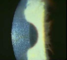
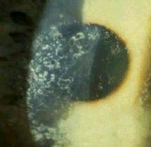
Fuch's Corneal Dystrophy

  • Common
  • Guttae along Descemet's
  • Loss of endothelial cells leads to corneal edema
  • Treat with NaCl/ointments and decreased IOP
Fuchs Endothelial Dystrophy, Misc-001 Guttae

Posterior Polymorphous Membranous Dystrophy

  • Endothelium becomes multilayered and develops epithelial features
  • Bilateral
  • Autosomal Dominant
  • Polymorphous opacities at Descemet's appear as vesicles or broad bands with irregular scalloped edges
  • Iris abnormalities
  • Stationary or slowly progressive
  • Periodic observation and glaucoma management
Posterior Polymorphous Dystrophy, Misc-001 \Posterior Polymorphous Dystrophy, Misc-002


Test your knowledge and Take The Quiz


All pages are Copyright ©2006 by Dennis H. Goldsberry, M.D., P.E.
Reproduction or archival of any protion of these pages is strictly prohibited except by express written permission
.